EXERCISE AND MUSCLES
The muscles are the body’s engines – all movement depends on muscular contraction and relaxation. The joints and limbs are powered by muscular contraction and the physiology of muscle action is both beautiful and elegant.
Muscle function
The muscles of movement are the skeletal muscles known also as voluntary or striated muscles. They have a main body, the belly of the muscle, and bony attachments which are very strong fibrous cables called tendons. Most movements depend on muscles contracting thus bending or straightening the joints which connect the bones to which the tendons are attached. A good example is the biceps muscle, attached to the upper arm, the humerus, and inserted into the forearm bones, the radius and ulna. Contracting the biceps bends the elbow joint which connects these bones.
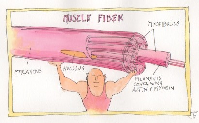
Each muscle is made up of thousands of fibres which in turn are made up of myofibrils. The myofibrils contain the microscopic structures which can bring about shortening of the whole structure – contraction. Each muscle cell contains thousands of myofibrils which are tubular structures extending the whole length of the cell. The myofibrils are made up of thick and thin filaments; the thick filaments made up mainly of myosin and the thin filaments of actin.
Muscle contraction is stimulated by discharge of the nerve supplying the muscle – the motor neuron (I won’t bore you with the physiology of this process. Suffice it to say that its effect is similar to the arrival of an electrical impulse). Each muscle is served by a large number of nerve fibres and the individual motor neuron plus the muscle fibres which it stimulates is called a motor unit. When an impulse reaches the muscle fibres of a motor unit, it stimulates a reaction in all the unit’s sarcomeres (the basic component of muscle) between the actin and myosin filaments. This reaction results in contraction or shortening of the muscle which is achieved by the myosin and actin filaments sliding alongside each other. Viewed under a microscopic the muscle fibres have a striped appearance – hence striated muscle – which is caused by the highly ordered arrangement of the actin and myosin within the fibrils and the similarly ordered arrangement of the fibrils within the muscle cell.
Muscle contraction continues until the neuron stops stimulating it or until muscle fatigue sets in. This happens when the supply of energy to fuel contraction is exhausted.
Muscle fibre types
One more important fact about muscles. There are two distinct types of fibre – slow twitch (Type I) and fast twitch (Type II) fibres (which are further subdivided into Type IIa and Type IIb fibres but please don’t trouble yourselves with that information). Slow twitch fibres contract slowly and can maintain their shortening for long periods – they are the strength and endurance giving fibres and are particularly used during such activities as distance running and cycling. Fast twitch fibres contract rapidly but tire quickly and are those most used by sprinters and weight lifters. Repeated contractions gives speed to action.
Most muscles consist of a combination of slow and fast twitch fibres but one often predominates in particular muscles – for instance back muscles which move little but contract continuously to maintain posture are predominantly slow twitch. Eye muscles on the other hand are almost all fast twitch. There is also a genetic influence on the predominance and distribution of muscle type. The ratio of slow to fast is about 50% for most of us. However some individuals inherit a much larger proportion of one or another and that may determine their prowess in different sporting endeavours. Those born with a higher proportion of Type I fibres are more likely to become endurance athletes while those born with more Type II fast twitch fibres will do better as sprinters.
Different exercise types use different proportions of slow (Type 1) and fast (Type 2) twitch fibres. Aerobic (with oxygen) exercise, like distance running, uses a predominance of Type 1 fibres which require a regular supply of oxygen plus other nutrients. Anaerobic (without oxygen) exercise involves short bursts of higher intensity contractions at a much greater percentage of their maximum contraction strength. Examples of anaerobic exercise include sprinting and weight lifting. It is fuelled by glucose and a compound called ATP which are quickly depleted so that anaerobic exercise cannot be sustained and fatigue soon sets in. However most exercises, like soccer, climbing and wood chopping are partially aerobic and partially anaerobic.
PS
Marathon running, blood vessels and knees
Some interesting news this week. You may have read about the study of the effects of training for marathon novices. 138 first time marathon completers were tested six months before the London Marathon and the tests were repeated soon after the race. The main findings were a reduction of blood pressure and a lessening of arterial stiffening. Both of these changes reduce the risk of future heart attacks or strokes. The older, slower runners benefited most.
These findings are not particularly surprising. Much more interesting, and much less widely reported, was the effect of the marathon training on the knees of the athletes. In a separate arm of the study 71 first timer marathoners had their knees subjected to MRI scanning six months before and two week’s after the race. The results showed a reduction in the “radiological score of damage in: subchondral bone marrow oedema in the condyles of the tibia and femur”. In other words the main joint surfaces were more resilient after the training than before. There was some deterioration in the cartilage of the knee cap, without any apparent symptoms. It appears, to the surprise of many, that running, even on hard surfaces, may be good for your knees. I will have lot more to say on this subject when I get to the complications of exercise in a few months time.
However I remain unenthusiastic about marathon running as a way of increasing or maintaining physical fitness and good health – it requires far more input than needed and does result in musculo-skeletal injuries. In the first part of the study there were 52 drop outs – 27% of those who were initially enrolled in the study. However it is clearly a lot better than doing nothing!
Subscribe to the blog
Categories
- Accelerometer
- Alzheimer's disease
- Blood pressure
- BMI
- Cancer
- Complications
- Coronary disease
- Cycling
- Dementia
- Diabetes
- Events
- Evidence
- Exercise promotion
- Frailty
- Healthspan
- Hearty News
- Hypertension
- Ill effects
- Infections
- Lifespan
- Lipids
- Lung disease
- Mental health
- Mental health
- Muscles
- Obesity
- Osteoporosis
- Oxygen uptake
- Parkinson's Disease
- Physical activity
- Physical fitness
- Pregnancy
- Running
- Sedentary behaviour
- Strength training
- Stroke
- Uncategorized
- Walking


